Home
Uncategories
Foot Muscles Mri : New Insights Into Intrinsic Foot Muscle Morphology And Composition Using Ultra High Field 7 Tesla Magnetic Resonance Imaging Bmc Musculoskeletal Disorders Full Text - The flexor digiti minimi brevis (flexor brevis minimi digiti, flexor digiti quinti brevis) lies under the metatarsal bone on the little toe, and resembles one of the interossei.
Foot Muscles Mri : New Insights Into Intrinsic Foot Muscle Morphology And Composition Using Ultra High Field 7 Tesla Magnetic Resonance Imaging Bmc Musculoskeletal Disorders Full Text - The flexor digiti minimi brevis (flexor brevis minimi digiti, flexor digiti quinti brevis) lies under the metatarsal bone on the little toe, and resembles one of the interossei.
Foot Muscles Mri : New Insights Into Intrinsic Foot Muscle Morphology And Composition Using Ultra High Field 7 Tesla Magnetic Resonance Imaging Bmc Musculoskeletal Disorders Full Text - The flexor digiti minimi brevis (flexor brevis minimi digiti, flexor digiti quinti brevis) lies under the metatarsal bone on the little toe, and resembles one of the interossei.. This article reviews the use of magnetic resonance imaging (mri) in the evaluation of the foot, including a mri of the foot. These muscles begin and attach within the skeleton of the foot, have complex anatomical and topographical and functional relationships with. ► hip ► pelvis ► thigh ► knee ► lower extremity/shin ► ankle ► foot. Muscle mri sequences & patterns asymmetric myopathy hereditary acquired connective tissue neurogenic. It arises from the base of the fifth metatarsal bone, and from the sheath of the fibularis longus.
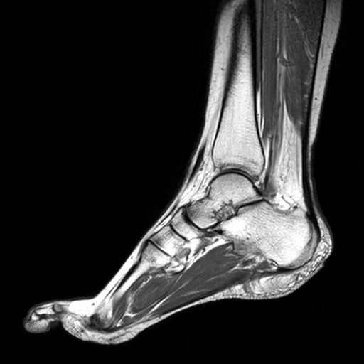
Bone contusions, osteonecrosis, marrow oedema syndromes, and stress > fractures) > synovial based disorders ( eg. Indications for foot mri scan. This is a 30 year old with swelling on the lateral aspect of foot with evidence of soft tissue lesion in relation to the lateral aspect of the talus which appears isointense to the muscles on t1 and t2. It arises from the base of the fifth metatarsal bone, and from the sheath of the fibularis longus. Lateral and medial processes of calcaneal tuberosity.
Mri Sliders Mri Anatomic Imaging Of The Foot Mr Tip Com from www.mr-tip.com It arises from the base of the fifth metatarsal bone, and from the sheath of the fibularis longus. In addition, an image of all the muscles of the back and. Learn more details about them at kenhub! Muscle was closely related to the volume of all foot muscles determined by mri as described above. Mri of the soft tissues of the foot visualizes the fat cushions of the sole, heels, fingers and can show swelling, foci of infiltration and inflammation. Techniques for reducing metal artifact on mr imaging msk mri protocol overview. Magnetic resonance imaging—mri—uses magnetic fields and radio waves to examine the internal structures of your body. Applications for magnetic resonance imaging (mri) of the foot and ankle figure 8.4 image planes for foot and ankle mri.
Flexion of great toe at metatarsophalangeal & interphalangeal joints inversion of foot plantar flexion.
Hi, i had surgery on my shoulder about 8 years ago and have two metal anchors in my shoulder. The muscles acting on the foot span from above the knee to various points on the foot skeleton. Mri with hardware in foot? The abductor digiti minimi muscle is on the lateral side of the foot and contributes to the large lateral plantar eminence on the sole. Upper and lower lines mark. ► hip ► pelvis ► thigh ► knee ► lower extremity/shin ► ankle ► foot. Mri with hardware in foot? Subscribe to foot & ankle problems. The flexor digiti minimi brevis (flexor brevis minimi digiti, flexor digiti quinti brevis) lies under the metatarsal bone on the little toe, and resembles one of the interossei. Lateral and medial processes of calcaneal tuberosity. A magnetic resonance imaging (mri) was performed on a normal subject; Learn about foot and ankle mri here. Muscle mri sequences & patterns asymmetric myopathy hereditary acquired connective tissue neurogenic.
Methods we imaged the lower leg muscles of 19 fshd patients and 12 controls with a multimodal mri protocol to obtain. .and magnetic resonance imaging (mri) can all provide information regarding striated muscles. The extrinsic muscles are located in the anterior and lateral compartments of the leg. Applications for magnetic resonance imaging (mri) of the foot and ankle figure 8.4 image planes for foot and ankle mri. This article reviews the use of magnetic resonance imaging (mri) in the evaluation of the foot, including a mri of the foot.
Ankle Tendons Topographic Anatomy Radiology Case Radiopaedia Org from prod-images-static.radiopaedia.org Muscles of the ankle and foot. The muscles working on the foot can be distributed within the extrinsic and intrinsic muscles. Indications for foot mri scan. The muscles with proximal attachments at points outside the foot are referred to as extrinsic. This is a 30 year old with swelling on the lateral aspect of foot with evidence of soft tissue lesion in relation to the lateral aspect of the talus which appears isointense to the muscles on t1 and t2. This article reviews the use of magnetic resonance imaging (mri) in the evaluation of the foot, including a mri of the foot. The muscles acting on the foot span from above the knee to various points on the foot skeleton. Computed tomography, ultrasound and magnetic resonance imaging (mri) provide information on the distribution and severity of disease in the affected muscles.
In addition, an image of all the muscles of the back and.
Subscribe to foot & ankle problems. Learn about foot and ankle mri here. Mri with hardware in foot? .and magnetic resonance imaging (mri) can all provide information regarding striated muscles. Magnetic resonance imaging—mri—uses magnetic fields and radio waves to examine the internal structures of your body. Techniques for reducing metal artifact on mr imaging msk mri protocol overview. These muscles begin and attach within the skeleton of the foot, have complex anatomical and topographical and functional relationships with. Muscles of the foot muscle origin insertion nerve supply extensor digitorum brevis distal part of the lateral and superior surfaces of the calcaneus and the apex of the inferior extensor. ► hip ► pelvis ► thigh ► knee ► lower extremity/shin ► ankle ► foot. Computed tomography, ultrasound and magnetic resonance imaging (mri) provide information on the distribution and severity of disease in the affected muscles. Learn more details about them at kenhub! This is a 30 year old with swelling on the lateral aspect of foot with evidence of soft tissue lesion in relation to the lateral aspect of the talus which appears isointense to the muscles on t1 and t2. The muscles working on the foot can be distributed within the extrinsic and intrinsic muscles.
Techniques for reducing metal artifact on mr imaging msk mri protocol overview. An overview of the intrinsic muscles of the foot including their origin, insertion, blood supply, innervation, function and clinical relevance. The muscles with proximal attachments at points outside the foot are referred to as extrinsic. Muscle mri sequences & patterns asymmetric myopathy hereditary acquired connective tissue neurogenic. Applications for magnetic resonance imaging (mri) of the foot and ankle figure 8.4 image planes for foot and ankle mri.
Radiological Anatomy X Ray Ct Mri Kenhub from thumbor.kenhub.com This article reviews the use of magnetic resonance imaging (mri) in the evaluation of the foot, including a mri of the foot. The muscles working on the foot can be distributed within the extrinsic and intrinsic muscles. Subscribe to foot & ankle problems. Human anatomy for muscle, reproductive, and skeleton. Explore more like foot muscle anatomy mri. A magnetic resonance imaging (mri) was performed on a normal subject; Magnetic resonance imaging—mri—uses magnetic fields and radio waves to examine the internal structures of your body. Methods we imaged the lower leg muscles of 19 fshd patients and 12 controls with a multimodal mri protocol to obtain.
.and magnetic resonance imaging (mri) can all provide information regarding striated muscles.
Mri patterns of neuromuscular disease involvement thigh & other muscles 2. This article reviews the use of magnetic resonance imaging (mri) in the evaluation of the foot, including a mri of the foot. Subscribe to foot & ankle problems. Explore more like foot muscle anatomy mri. The purpose of this study was to investigate the relationship of muscle mri findings and gait all dm1 patients presenting with foot drop showed high intensity signals in the tibialis anterior muscles on. Lateral and medial processes of calcaneal tuberosity. ► hip ► pelvis ► thigh ► knee ► lower extremity/shin ► ankle ► foot. Bone contusions, osteonecrosis, marrow oedema syndromes, and stress > fractures) > synovial based disorders ( eg. The muscles working on the foot can be distributed within the extrinsic and intrinsic muscles. Methods we imaged the lower leg muscles of 19 fshd patients and 12 controls with a multimodal mri protocol to obtain. Muscles of the ankle and foot. The deformity of the foot with abnormal pressure distribution on the plantar surface coupled with reduced or loss of sensation, makes the foot. Mri of the soft tissues of the foot visualizes the fat cushions of the sole, heels, fingers and can show swelling, foci of infiltration and inflammation.

0 Comments:
Posting Komentar